|
IV. – A COMPLETE MOSASAUR SKELETON,
OSSEOUS AND CARTILAGINOUS.
___________________________________________________
By Henry Fairfield Osborn
Plates XXI, XXII, and XXIII.
____________________________________
In the spring of 1898, Prof. S. W. Williston's fine memoir upon the Kansas
Mosasaurs seemed to cover the subject completely, summing up all the facts derived from
the great Kansas University collection, as well as many of the results of the labors of
Cuvier, Owen, Marsh, Cope, Dollo, Baur, and others. But it appears impossible to say the
last word in palaeontology. Professor Williston himself has recently described a portion
of the nuchal fringe of Platecarpus, as well as the epidermal fin
contours. The remarkable specimen which forms the subject of the present brief memoir
throws new and welcome light not only upon Tylosaurus
but upon the anatomy of the Mosasaurs in general.
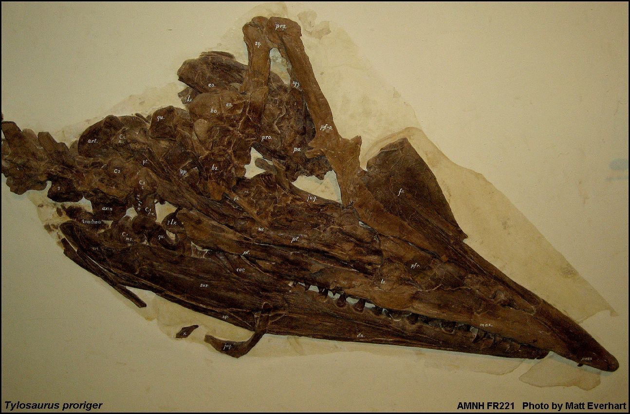
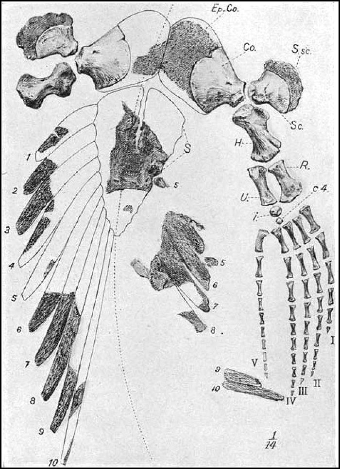
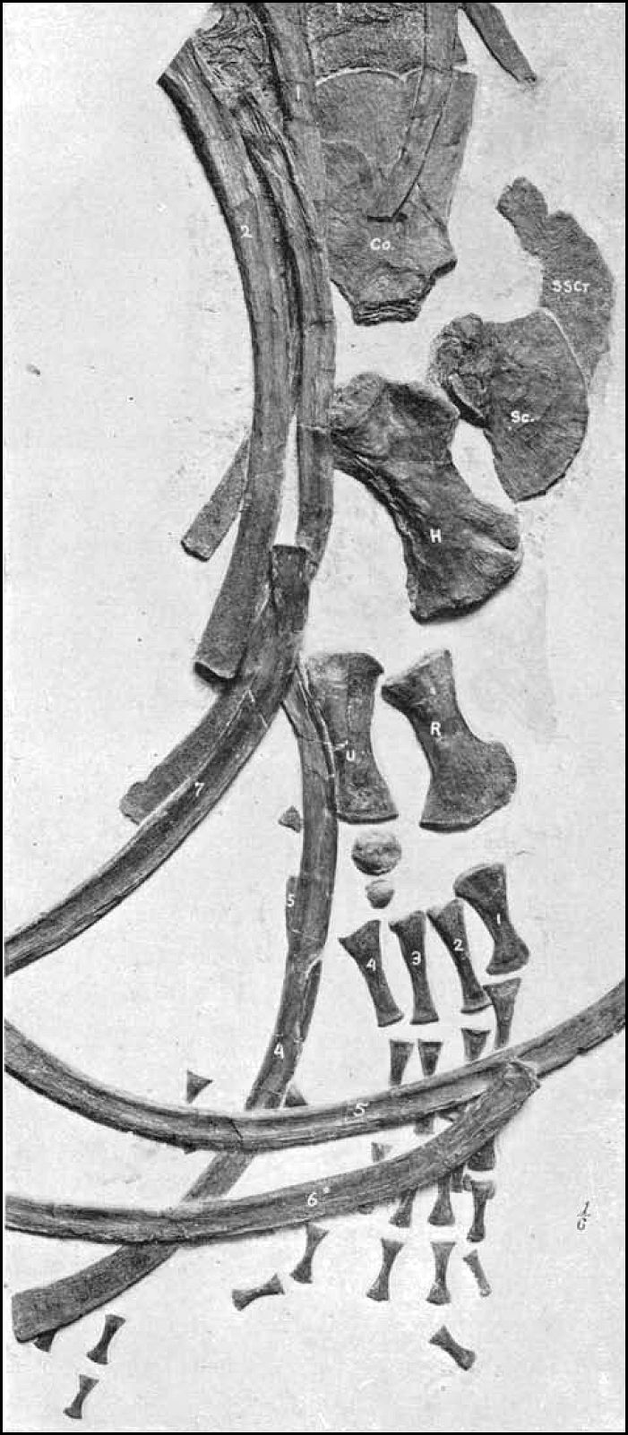
Together with the practically complete bony skeleton, the chief feature is the
unique preservation of the cartilages of the throat and chest, portions of the larynx,
trachea, bronchi, the epicoracoids, as well as the suprascapulć, the sternum, and sternal
ribs. Originally these parts were preserved entire, and we must deeply regret that before
this specimen came into possession of the Museum, much damage was done to the relatively
inconspicuous cartilages, in course of removal of the bones. Nevertheless 'Mr. Bourne, of
Scott City, Kansas, who excavated the fossil, deserves great credit for the skill and care
with which the conspicuous parts were removed.1
1 The specimen was examined before its purchase by
Dr. W. D. Matthew, and packed for shipment by Mr. Handel T. Martin. At the time no one
could judge of the existence of the cartilages or of the exceptionally complete condition
of the skeleton.
[167] |
168
OSBORN, A COMPLETE MOSASAUR SKELETON.
The specimen reached the Museum in a series of large slabs of Kansas chalk, and was
worked out under the direction of Mr. Hermann, by Mr. Thompson. With one exception, all
the contours of the original slabs were preserved and fitted together by their edges, as
in the original bedding; therefore the animal with all its parts, excepting a few minor
pieces, lies exactly as it was imbedded in the rocks. The original matrix surrounds
practically all the bones, and can be distinguished from the buff-colored outlying
plaster, even in the photographs, by its somewhat darker shades. The whole is mounted upon
a panel twenty-five feet long and permanently placed in a corridor which is to be devoted
to marine reptiles.
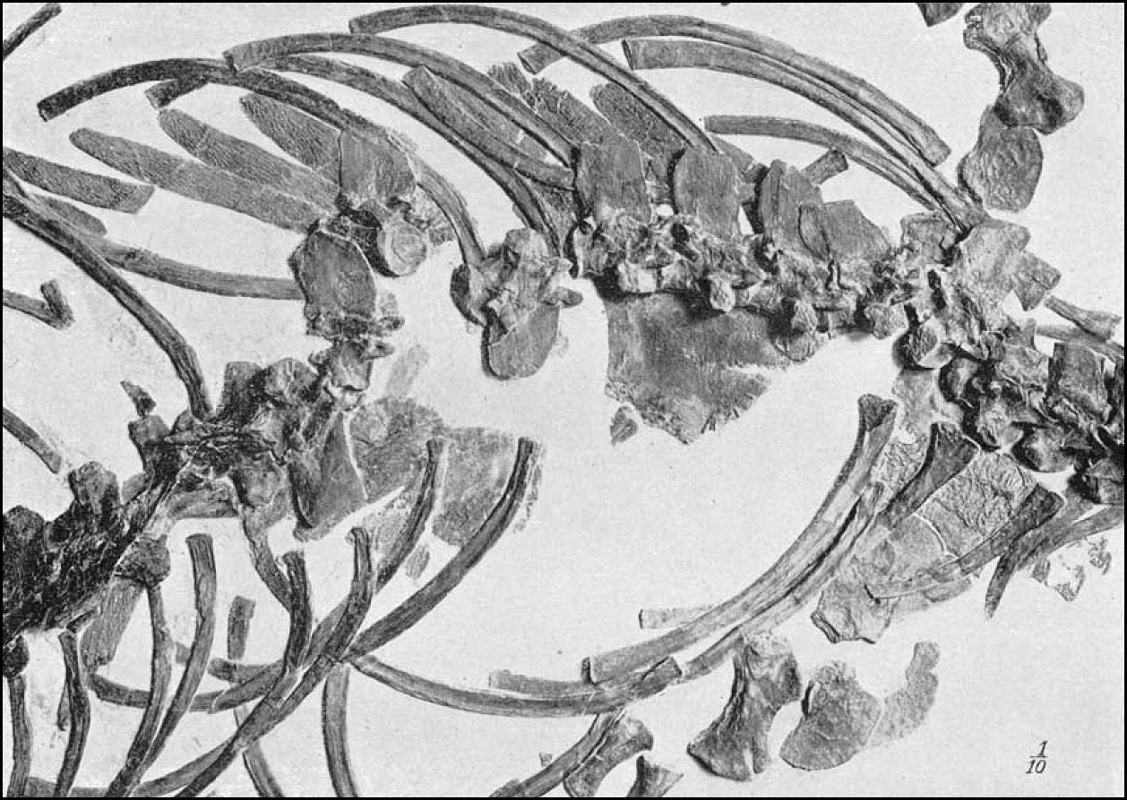
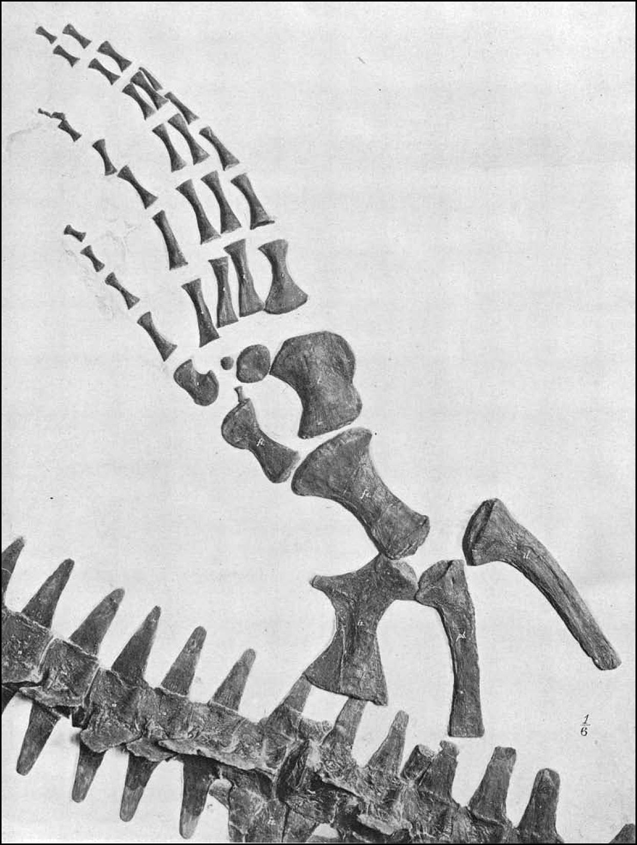
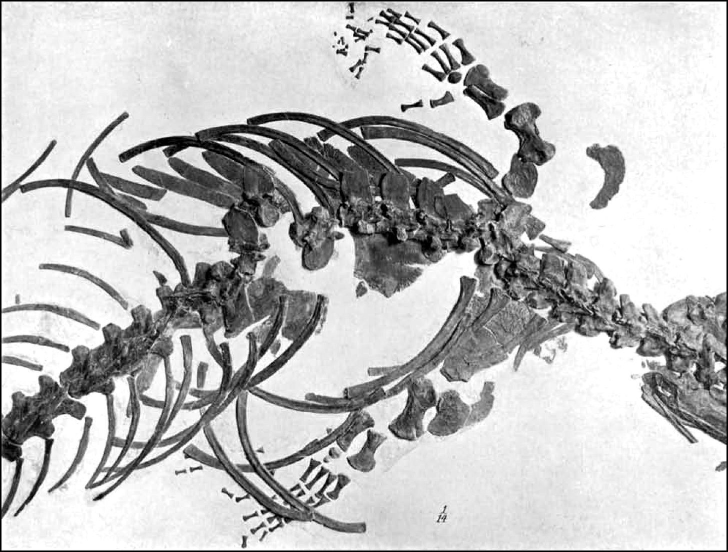
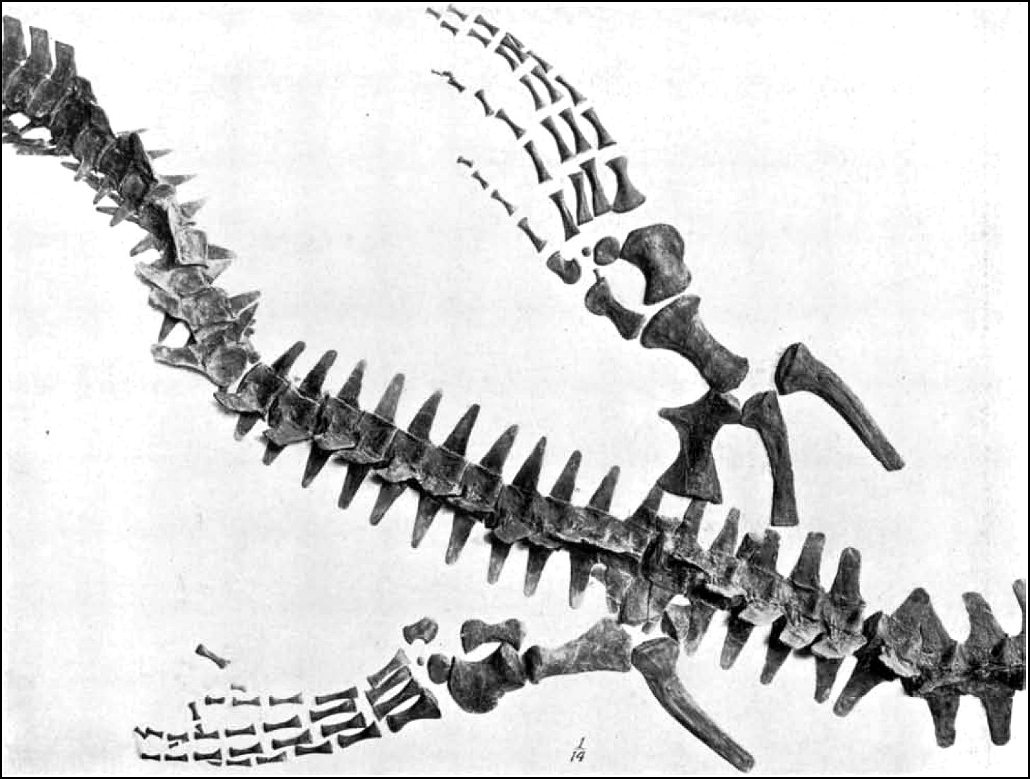
Position of the Skeleton. -The animal
lies outstretched upon its ventral surface, so that all the bones are exposed upon the
dorsal or lateral surfaces, excepting the left humerus and ulna, which are overturned. The
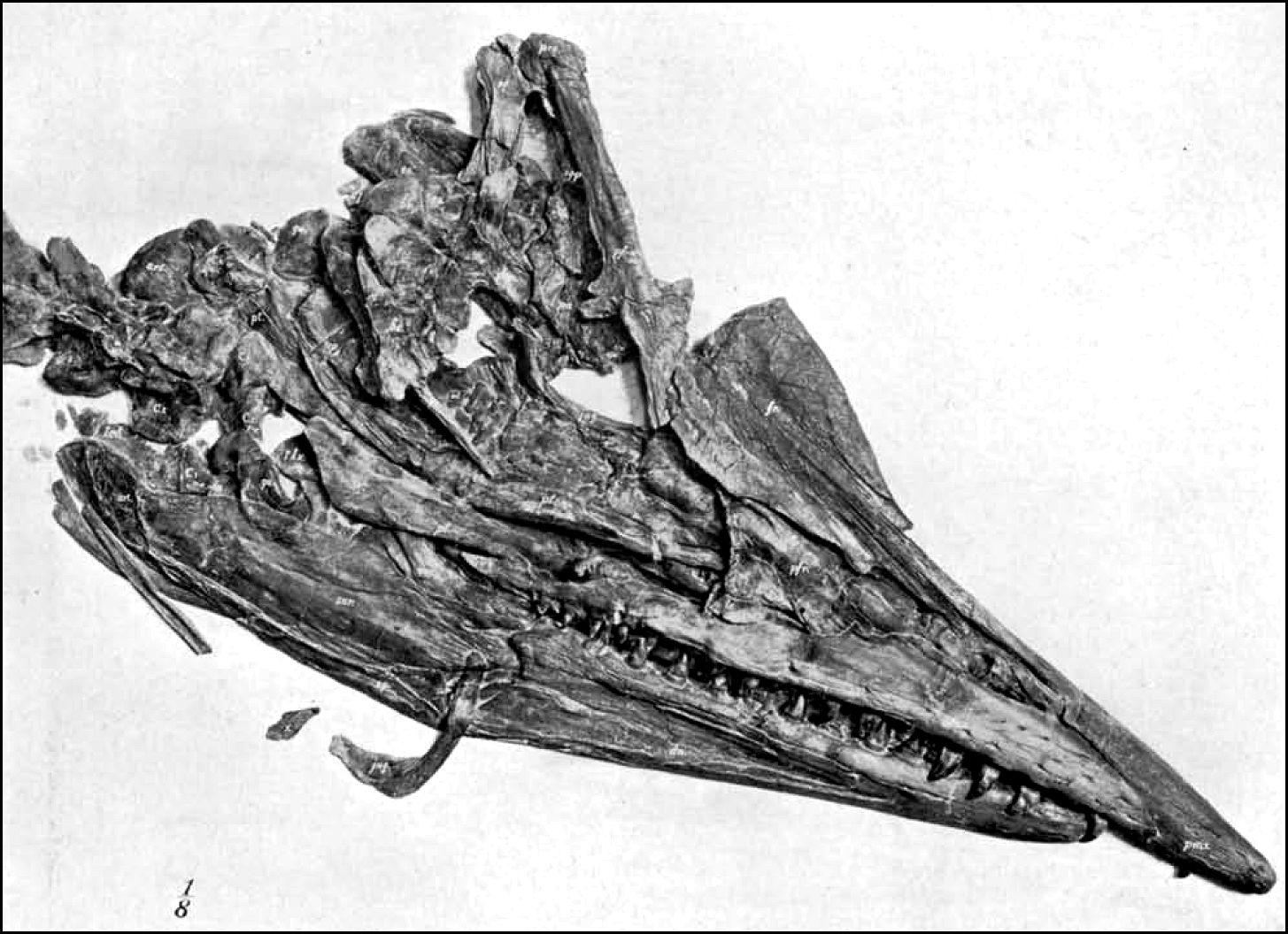
skull is crushed to
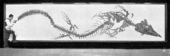
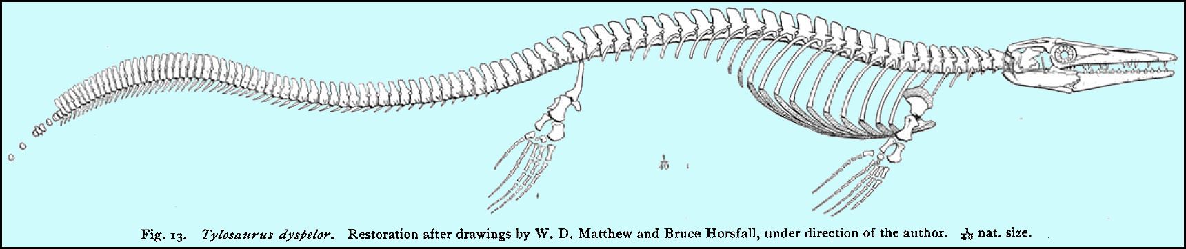
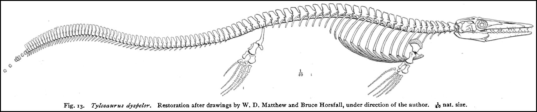
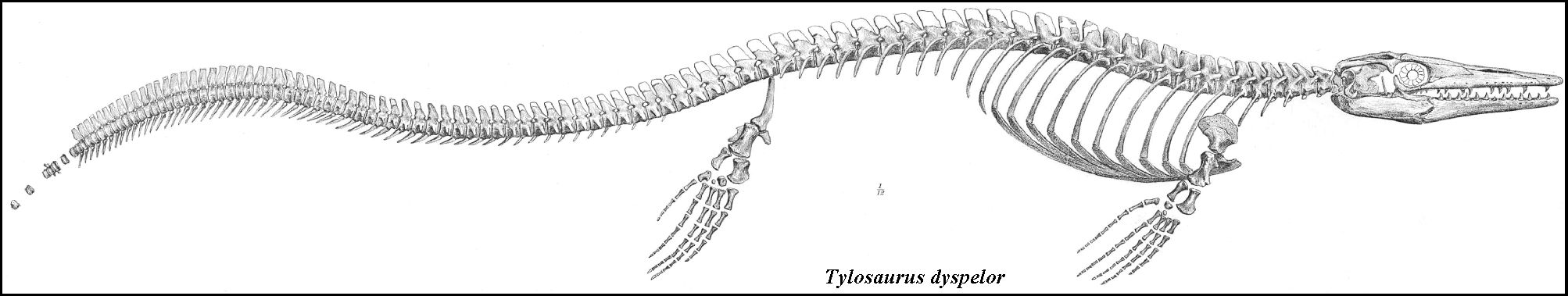
Fig.
I. Complete skeleton of Tylosaurus dyspelor,
in frame. 1/54 nat. size.
the left, together with the vertebrae, as far back as the 6th
dorsal. From the 7th to the 10th dorsals the
vertebrae are confused and displaced. The 11th dorsal to 29th caudal are horizontal with
the transverse processes outspread and the spines crushed to the right and left. The
remaining caudals, 30th-70th, lie upon the left side apparently in a natural position. The
pelvis and hind paddles have evidently shifted backwards in settling, so that the mooted
question of the position of the sacral vertebra cannot be positively settled by this
specimen.
Examination and Restoration. - The study
of the animal has been cooperative. Dr. W.- D. Matthew has carefully examined several
regions, and made a number of original suggestions and valuable criticisms in points of
interpretation, especially as to the remarkable tail curvature, the atlas complex, and the
general structure of the vertebrae. Dr. J. H. McGregor has greatly aided the writer in
studying and restoring the sternal region. The photographs are the work of Mr. A. E.
Anderson. The drawings are by Mr. Bruce Horsfall. |
OSBORN, A COMPLETE MOSASAUR SKELETON.
169
Tylosaurus dyspelor Cope.
[see note above]
Specific Characters. -This specimen
agrees very closely in size with Cope's cotype of T.
(Liodon) dyspelor,
found in 1871 at Fort Wallace, Kansas, and described by him in the 'Cretaceous Vertebrata'
(p. 167). The skull agrees exactly in size with the fine one mounted in the Munich Museum,
described by Merriam (1894, Taf. II) as T. proriger.
Size is no criterion, or at best an uncertain criterion of a species, but Williston
advances (1898, p. 175) no other satisfactory means of separating T. dyspelor from T. proriger. Thirty-five feet is the length
assigned by this author to the largest Tylosaurs, a length considerably exceeding that of
the present specimen. It is evident that a young T.
dyspelor might exhibit exactly the measurements of T. proriger.
We observe, however, in this specimen certain characters which may possibly prove
to specifically distinguish this type from T. proriger, as follows:
1. 22 dorsals. Williston assigns 23 dorsals to T. proriger.
2. No rib upon the axis. A rib if present upon C.3 was certainly very small.
Williston figures ribs upon both axis and C.3.
3. A dorsal curvature of the mid-region of the tail, not observed in T. proriger.
4.
Phalanges in the manus estimated at 39. In T. proriger, same estimated at 47.
None of these characters, however, are absolutely determined in both types, so as
to be clearly distinguishable. A summary of the chief anatomical characters is given in
the conclusion.
Measurements and Proportions.
WITH
SLIGHT CORRECTIONS FOR CRUSHING.
Eng. Meas. Metres.
Skull, from back of supratemporal arch to rostrum……....................…
3’ 11” 1.19
Jaw, angle to rostrum,
approximate……………………….................
…..3’ 10”
1.16
Seven cervical vertebrae,
actual…………………….....…............….….
1’ 11”
.58
Twenty-two dorsal
vertebrae,actual…………………….................……. 7’ 11” 2.41
Ten dorsals, with sternal ribs, actual…………………..
……............... 3’ 7” 1.09
Twelve dorsals, with floating ribs,
actual………………........................ 4’ 11”
1.32
Seventy caudals and pygals, actual length, as mounted (including
spaces left for eight intermediate caudals towards extremity of tail)
13’ 9”
4.20
Total length of tail, estimated
……………………...............…………… 15’ 4.57
Fore paddle from head of humerus to tip, estimated………................…... 2’ 11”
.90
Hind paddle from head of femur to
tip……………………….................… 3’ 3” .98
Total length from tip of rostrum to last, or 78th, caudal, as mounted .. 27’ 4”
8.34
According to Williston the tail terminates very abruptly in Tylosaurus proriger, in contrast with its gradual
and slender termination in Platecarpus, as
described below. If this was the case in this specimen of T.
dyspelor, we |
170
OSBORN, A COMPLETE MOSASAUR SKELETON
should not allow more than 15 inches or 38 centimetres
additional, giving us a total length of about 29 feet or 8.83 metres.
The proportions of different regions of the body, as Williston has shown, are very
characteristic of different genera of Mosasaurs. In this individual the total of 29 feet
or 9 metres is roughly distributed as follows:
Feet.
Metres.
Head and
jaw……………………………
4 1.22
Neck…………………………………….
2 .61
Back
……………………………………
8 2.44
Tail
….………………………………….
15 4.56
Total…………………………… 29
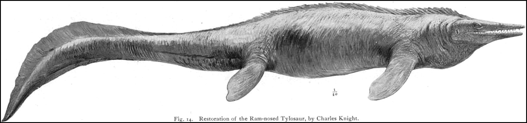
Thus the back is four times the length of the neck, twice the length of the head,
and about one half the length of the tail. In other words, the tail is longer than the
other regions of the body combined. These proportions are carefully observed in Mr.
Knight's restoration.
|
