skull-8b.jpg (90211 bytes)  

Platecarpus tympaniticus

A mosasaur from the upper Smoky Hill Chalk of western Kansas


Copyright © 2000-2009 by Mike Everhart

last update 09/16/2009

LEFT: View of skull and lower jaws of FHSM VP-17017 as found. Skul was upside down and lower jaws were separated and lying off to the side.

IMPORTANT NOTE:  The recent publication of an article in the JVP by Takuya Konishi and Michael Caldwell clarifying the identification and relationships of the various species of Platecarpus will necessitate some major changes in some of my web pages. Please note that Platecarpus planifrons Cope (1874) is now identified as the most common species of Platecarpus 
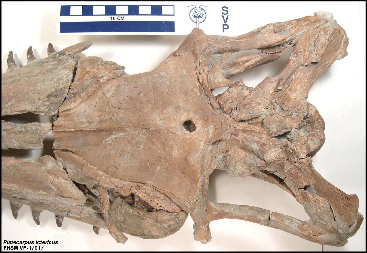
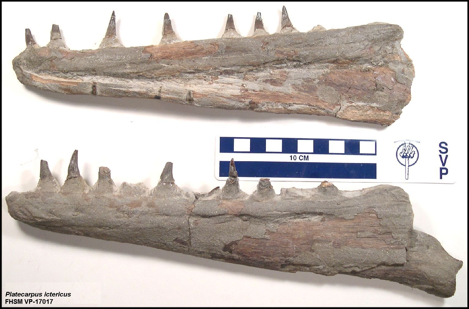
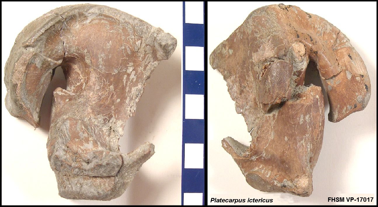
in the lower chalk (late Coniacian to middle Santonian), and P. ictericus (Cope, 1871) is the most common species of this genera in upper chalk (middle Santonian through early Campanian). P. coryphaeus (Cope, 1872) is a junior synonym of P. ictericus.  The name Platecarpus tympaniticus (Cope, 1869) is now limited to a single specimen (holotype) from Mississippi. The species that I had previously identified as Platecarpus planifrons (above) is now "unidentified" and possibly a new genus / species which we are working to identify / describe.  I consider this paper to be a major improvement in mosasaur phylogeny. The citation is:

Konishi, T. and Caldwell, M. W. 2007. New specimens of Platecarpus planifrons (Cope, 1874) (Squamata: Mosasauridae) and a revised taxonomy of the genus: Journal of Vertebrate Paleontology 27(1): 59-72. 

And another revision.. Platecarpus tympaniticus is now the senior synonym over P. ictericus and P. coryphaeus. 

Konishi, T., Caldwell , M.J. and Bell , G.L., Jr. 2010. Redescription of the holotype of Platecarpus tympaniticus Cope 1869 (Mosasauridae: Plioplatecarpinae), and its implications for the alpha taxonomy of the genus. Journal of Vertebrate Paleontology 30(5):1410-1421. 

This Platecarpus tympaniticus  mosasaur skull was found and mostly prepared in 1996, but was not photographed in any detail until January, 2000.

craniu1a.jpg (3776 bytes) A dorsal view of the back of the mosasaur skull. Scale is 15 cm (about 6 inches).
frontala.jpg (3236 bytes) A slightly closer view of the frontal bone which covers most the top of the skull. The parietal, parietal foremen and parietal-frontal suture are also visible.
pof-suta.jpg (3683 bytes) A close up the suture between the frontal and the post orbital frontal (POF).  The POF provides the upper and rear portion of the orbit of the eye.  The jugal and prefrontal complete the rest of the orbit. The large round object at the top of the picture is the left quadrate bone. For some unknown reason, both quadrates had been displaced forward into the orbits before the skull was buried.
craniu2a.jpg (4148 bytes) A ventral view of the back of the skull, showing the left pterygoid bone and the displaced left quadrate.  The rounded surface at the far right is the occipital condyle (where the vertebral column is attached to the skull).
occ-cona.jpg (4101 bytes) The back of the skull from slightly above. The occipital condyle is in the center of this picture.
muzzle1a.jpg (3703 bytes) A dorsal view of the snout or muzzle of the mosasaur.  The premaxilla is centered between the right and left maxillaries.  The nasal openings (nares) of the skull are located near the center of the picture.
muzzle2a.jpg (3987 bytes) A ventral view of the muzzle.   There were four teeth in the premaxilla and twelve teeth in each of the maxillaries..
premax2a.jpg (3200 bytes) A close-up, ventral view of the premaxilla and anterior portions of both maxillaries.
quad-ra.jpg (2825 bytes) A lateral view of the right quadrate.  The right eardrum of the mosasaur was located on this surface.
quad-r2a.jpg (2544 bytes) A medial view of the right quadrate.  The cracks were probably the result of extreme pressures during fossilization.
quad-la.jpg (3228 bytes) A medial view of the left quadrate.   The broken bones at the upper left and lower right are probably the ends of the jugal which forms the back and lower portions of the orbit of the left eye.
quad-l2a.jpg (4722 bytes) A wider view of the left quadrate and the left pterygoid. 
dentarya.jpg (4731 bytes) Lateral views of the right (top) and left (bottom) lower jaws. The jaws were found laying together and detached from the rest of the skull.
dentar2a.jpg (4679 bytes) Medial views of the right (top) and left (bottom) dentarys. There are twelve teeth on each of the dentarys.
teetha.jpg (3184 bytes) Loose teeth that were found associated with the specimen.  These were probably new (forming) teeth that were not yet set firmly in the jaws.
skull2a.jpg (2744 bytes) An overhead view of the complete skull, lower jaws and cervical vertebrae.
cer-vera.jpg (3706 bytes) Four cervical vertebrae in articulation as found.
atlasaxa.jpg (2807 bytes) The re-assembled atlas-axis vertebrae complex.   In mosasaurs, the atlas vertebra is made up of four pieces of bone that articulate between the occipital condyle of the braincase and the axis vertebra.
bitesa.jpg (2963 bytes) Serrated bite marks on the lower edge of the left jaw are evidence of scavenging by Squalicorax sharks.  Many of the mosasaur's ribs had similar bite marks.  There was no limb material found with this specimen, suggesting that the carcass had been stripped fairly completely before it sank to the bottom of the seaway. In 1999, a large Squalicorax kaupi tooth was found at the same locality.
bites2a.jpg (2998 bytes) More shark bite marks on the angular of the left jaw.

LARGER, UPDATED  PICTURES

FHSM VP17017Aa.jpg (30715 bytes) LEFT: The skull of FHSM VP-17017 in dorsal view. The teeth would normally point downward, not off to the side.
FHSM VP17017Ba.jpg (22456 bytes) LEFT: Close up of the back of the skull of FHSM VP-17017 in dorsal view
FHSM VP17017Ca.jpg (19635 bytes) LEFT: Right (top) and left dentarys of FHSM VP-17017 in medial and lateral views
FHSM VP17017Da.jpg (26514 bytes) LEFT: The right quadrate of FHSM VP-17017 in lateral and medial views.

LINKS

Click here to see the updated pictures from the 1996 dig for this mosasaur.

Click here to see other Platecarpus mosasaur remains at the Virtual Mosasaur Museum

Click here to see the mosasaurs at the Sternberg Museum of Natural History

Click here to see Samuel Williston's Platecarpus drawings published in 1898