CIMOLIASAURUS
25
...(previous text omitted)...
CIMOLIASAURUS.
Cimoliasaurus
magnus.
Cimoliasaurus magnus, LEIDY, Proc. Acad.
Nat. Sci. Phila. 1851, 325; 1854, 72, pl. ii, figs. 4, 5, 6.
Vertebræ differing from any of those
described in the preceding pages, and belonging to a huge Saurian, are frequently found in
the Greensand deposits of
4
March, 1865.
26
CIMOLIASAURUS.
New Jersey. The vertebræ have slightly biconcave bodies and are usually well
preserved, though all the specimens I have had the opportunity of examining have had their
arches and processes broken off, apparently after their discovery. Of such vertebræ
thirteen specimens, from Burlington County, N. J., were presented to the Academy of
Natural Sciences by Dr. S. G. Morton. They appear to have belonged to the same individual,
and consist, as I suppose them to be, of two dorsals and eleven lumbars.
The body of the dorsal vertebræ, Figs.
13-16, Plate V, is the transverse section of a cylinder compressed from above downward and
contracted towards the middle, resembling in form the body of the cervical vertebra
referred to Discosaurus. The articular faces are transversely oval, slightly
emarginate above, and are more concave than in the cervical vertebra of Discosaurus.
They present a central prominence, are beveled off at the border, and are defined by a
subacute edge from the rest of the body. A pair of large venous foramina underneath the
latter communicate with channels opening by a single large orifice in the spinal canal,
which is depressed towards the middle and wide. The vertebral arch has been coossified
with the body, but its loss prevents me from ascertaining anything in regard to its form.
In one of tile specimens, as represented in figs, 13, 14, 15, there projects from the
middle of the side of the body a short, robust, cylindroid transverse process, terminating
in a large irregular facet for the articulation of a rib. In the other vertebra, Fig. 16,
probably a more anterior one, the transverse process is broken, but its base indicates it
to have been of greater vertical extent than in the former specimen, though not quite so
wide, nor does it extend so low, but above appears nearly to have reached the abutment of
the vertebral arch.
The size of the two specimens is nearly equal, their
measurements being as follows:
Lines.
Length of the vertebral body
32-33 [ 6.7 cm]
Breadth of articular surfaces
52 [10.9 cm]
Height
42 [ 8.8 cm]
Width of spinal canal at the middle
12 [ 2.5 cm]
Width of spinal canal at the extremities
15 [ 3.2 cm]
Breadth between articular facets of transverse processes
72 [15.1 cm]
The eleven lumbar vertebræ, of which the largest and
smallest specimens are represented in Figs. 17-19, Plate V, and 16-18, Plate VI, do
not form an unbroken series, but the specimens successively diminish in size from nearly
that of the dorsals just described, to a size rather less than the supposed caudals of Discosaurus.
They are nearly identical in form throughout, The more anterior have the body absolutely
somewhat longer than in the dorsals, though the other diameters are diminished. The
articular extremities are also slightly more dished than in the dorsals, and almost devoid
of the central prominence. The venous foramina on the under part of the body are nearer
together, and the intervening portion of bone appears pinched into a convex ridge, The
more posterior specimens, which may be regarded as caudals, have the articular extremities
of their body rather more concave, and underneath they do not form so prominent a ridge
between the position of the two venous foramina.
CIMOLIASAURUS.
27
In all the lumbar vertebræ the abutments of the
vertebral arch are fully coossified with their body, leaving no well-marked trace of the
former sutural connection. The spinal canal is wide, and is depressed at the floor towards
the middle, where it exhibits one or two large venous foramina. In all the specimens the
transverse processes have been broken off, but in all, their remaining bases are seen
projecting from the lower part of the side of the body. They spring from nearly the whole
width of the body, with which they were completely coossified, though they present the
appearance as if they formerly possessed a sutural attachment. They were evidently robust
and strong, and were directed obliquely outward and downward.
The sides of the body of the vertebræ form, together
with the sides of the vertebral arch and the upper part of the transverse processes, a
nearly uniform slope, broken only by a slight elevation formed by the apparent sutural
coossification of the transverse process with the body. The under part of the body between
the transverse processes nearly forms a level surface, more or less elevated into a ridge
between the venous foramina, and depressed along a line with the position of the latter.
Measurements, derived from the largest and the smallest
of the series of eleven lumbars, are as follows: -
LARGEST.
SMALLEST.
Lines.
Lines.
Length of body
35 24
[7.4-5.0 cm]
Breadth of articular surfaces
44 31
[9.2-6.5 cm]
Height of articular surfaces
36 22
[7.6-4.6 cm]
Width of spinal canal.
10
8 [2.1-1.7 cm]
The vertebræ, above described, were briefly noticed a
few years ago in the Proceedings of the Academy of Natural Sciences, volume V, page 325,
and referred to a Reptile under the name of Cimoliasaurus magnus.
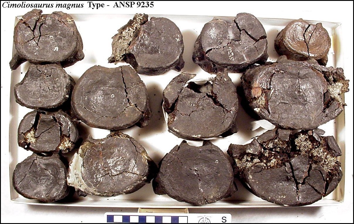
Fourteen vertebræ of the same Saurian as the preceding
have been submitted to my inspection by Mr. 0. R. Willis, through Prof. Cook. They all
evidently belonged to the same individual, and were obtained from the Green-sand, near
Freehold, Monmouth Co., N. J. Six of the specimens are dorsal, the remainder lumbar
vertebræ. Of the former, three appear to have had their transverse processes at the
conjunction of the vertebral arch and body; the others had them situated successively
lower on the sides of the body.
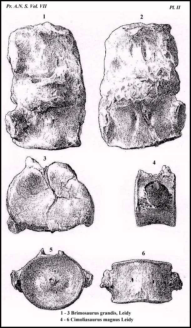
Two of the more anterior dorsals are represented in Figs.
1, 2, 3, 4, Plate VI. They exhibit a slight want of symmetry, which is the case also with
another anterior dorsal, but this character is a deformity, or mere individual
peculiarity. The body is a little longer and higher, in relation with the breadth, than in
the dorsals above described, and hence presents a more cylindrical form. The articular
extremities are moderately dished, and have a somewhat prominent annular margin. They are
nearly circular, but notched above, and are sharply defined by a subacute ridge from the
rest of the body. The bottom and sides of the latter are narrowed towards the middle or
are concave longitudinally, and they present a number of foramina, varying in size, which
communicate with venous channels opening into
28
CIMOLIASAURUS.
the spinal canal. The broken abutments of the vertebral arch are much broader and
stronger, in accordance with their being required to sustain the transverse processes,
than in the more posterior dorsals. The spinal canal is large and depressed towards the
middle of the floor. The measurements of one of the anterior dorsals, which are nearly of
the same size, are as follows: -
Lines.
Length of the vertebral body.
36 [ 7.6 cm]
Breadth of articular surfaces.
51 [10.7 cm]
Height of articular surfaces.
45 [ 9.5 cm]
Width of spinal canal.
12 [ 2.5 cm]
A more posterior dorsal vertebra, represented in Fig. 5,
Plate VI, differs from the preceding in the less length and depth of the body and the
slightly greater breadth, but chiefly in the lower position of the transverse process,
which extends from the vertebral arch to near the middle of the side of the body. The
measurements of this specimen are as follows: -
Lines.
Length of the vertebral body
33 [ 6.9 cm]
Breadth of the articular surfaces.
54 [11.3 cm]
Height of the articular surfaces.
43 [ 9.0 cm]
The remaining two posterior dorsal vertebræ, represented
in Figs. 6-9, Plate VI, appear to be from near the termination of the series. They have
the same form of body as the Burlington County specimens of posterior dorsals, above
described, with which they also nearly agree in size. The transverse processes are short,
robust, irregularly cylindroid protuberances, projecting from the lower part of the side
of the body and terminating in an articulating facet for a rib. In-the foremost of the two
vertebræ, Fig. 7, the facets are sub-circular and irregularly convex; in the other, Fig.
9, they are transversely oval and irregularly concave. On the under surface of the body of
the former, Fig. 6, there are two large foramina on each side communicating with venous
channels opening into the spinal canal; in the latter, Fig. 8, the under part of the body
presents two very large venous foramina, between which the bone forms a convex ridge, not
existing in the preceding vertebra. Measurements of the two posterior dorsal vertebræ are
as follows: -
Lines. Lines.
Length of the body
33 35
[ 6.9- 7.4 cm]
Breadth of the body.
56 52
[11.8-10.9 cm]
Height of the body
40 [ 8.4 cm]
Width of spinal canal.
11 [ 2.3 cm]
Vertical diameter of facet for the rib.
18 16
[ 3.8- 4.0 cm]
Transverse diameter of facet for the rib.
18
20
[ 3.8- 4.2 cm]
The eight lumbar vertebræ, of which the largest and
smallest specimens are represented in Figs. 10-15, Plate VI, form a nearly unbroken
series, and followed close after the dorsal specimens just described. They correspond in
form and constitution with the Burlington County specimens, except that the median part of
their body beneath, between the position of the venous foramina, forms a more prominent
ridge.
CIMOLIASAURUS.
29
The measurements of the largest and smallest specimens,
or the first and last of the series, are as follows: -
LARGEST. SMALLEST.
Lines.
Lines.
Length of the vertebral body.
35
31 [ 7.4-6.5 cm]
Breadth of articular extremities.
50
41 [10.5-8.6 cm]
Height of articular extremities.
39
28 [ 8.2-5.9 cm]
Width of spinal canal
9
8 [ 1.9-1.7 cm]
It is probable that the vertebræ, above described as
lumbars, may be regarded in part as representing sacrals and caudals. Both dorsals and
lumbars bear some resemblance to the corresponding vertebræ of Cetaceans, except that in
these the transverse processes project from the middle of the sides of the body of the
lumbars instead of the lower part. The long series of vertebræ of Cimoliasaurus consisting
of lumbars apparently gradually merging into caudals, perhaps indicate the absence of a
true sacrum and posterior extremities, as in Cetaceans.
I cannot avoid the suspicion that the specimens referred
to Cimoliasaurus magnus do not belong to the same great reptile as those considered
as characteristic of Discosaurus vetustus. The supposed caudals of the latter I
have suspected to be anterior cervicals notwithstanding the apparent provision for the
articulation of chevron bones. If all the vertebral specimens be viewed as belonging to
one animal, they represent cervicals, dorsals, and lumbars of Discosaurus,
otherwise they represent a cervical and caudals of the latter, and dorsals and lumbars of Cimoliasaurus.
The vertebræ described as caudals of Discosaurus have almost the same size and nearly the
same form as the smaller lumbars or caudals attributed to Cimoliasaurus. A rib of
proportionate size, coossified with the costal pit in the former, would give them a
striking resemblance to the latter, except that in Cimoliasaurus the costal or
transverse processes project from the lower part of the sides of the body, whereas in Discosaurus
the costal pits are situated at the middle of the sides of the body. The vertebræ,
however, differ in other important particulars. Besides the absence of the conspicuous
inflections (supposed to have been intended to accommodate chevron bones in the caudals of
Cimoliasaurus the body beneath is nearly level between the transverse processes,
while in Discosaurus it is strongly convex in the corresponding position.
No portions of the skull nor specimens of teeth have been
discovered which, with any probability, could be referred either to Discosaurus or Cimoliasaurus.
|
| 126
REFERENCES T0 THE PLATES.
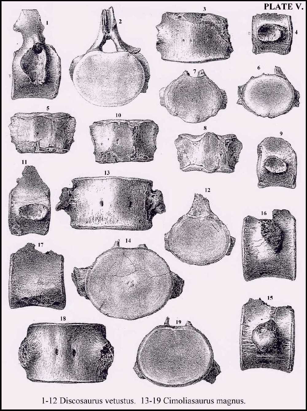
PLATE V.
All the figures are reduced one-half the diameter of the original specimens.
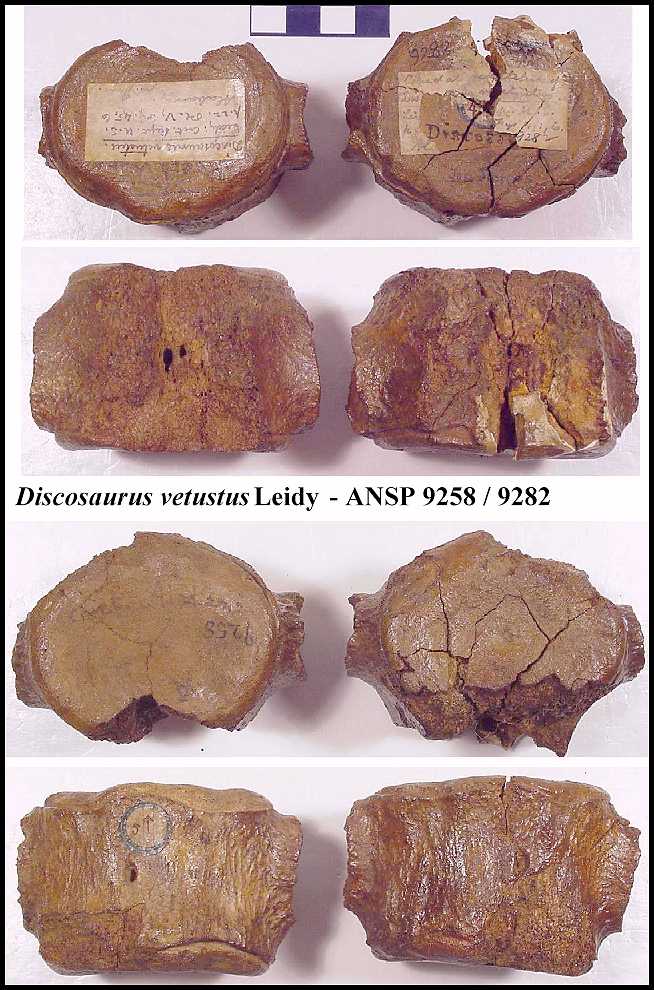
Figs. 1-12. Vertebræ of Discosaurus vetustus.
Figs. 13-19. Vertebræ of Cimoliasaurus magnus.
Figs. 1-3. Cervical vertebra of Discosaurus vetustus from Burlington
County, New Jersey.
Fig. 1. Lateral view; a, anterior surface of body; p, posterior
surface; c, costal pit.
Fig. 2. Posterior view. .
Fig. 3. Inferior view; the upper part is the anterior.
Figs. 4-6. Two caudal ? vertebræ of Discosaurus, from Alabama.
Fig. 4. Lateral view. a, costal pit.
Fig. 5. Inferior view of the same specimen.
Fig. 6. End view of a second specimen.
Figs. 7-9. Caudal ? vertebra of Discosaurus, from Burlington County, New
Jersey, found with the specimen above indicated from the same locality.
Fig. 7. End view.
Fig. 8. Inferior view.
Fig. 9. Lateral view.
Figs. 10-12. Caudal ? vertebra of Discosaurus, from Mississippi.
Fig. 10. Inferior view.
Fig. 11. Lateral view.
Fig. 12. End view.
Figs. 13-16, Dorsal vertebræ of Cimoliasaurus from Burlington County,
New Jersey.
Fig, 13. Inferior view.
Fig. 14. End view of the same specimen.
Fig. 15. .Lateral view of the same.
Fig. 16. Lateral view of a more anterior dorsal vertebra.
Figs. 17-19. A lumbar vertebra found with the preceding dorsals.
Fig. 17. Lateral view.
Fig. 18. Inferior view.
Fig. 19. End view.
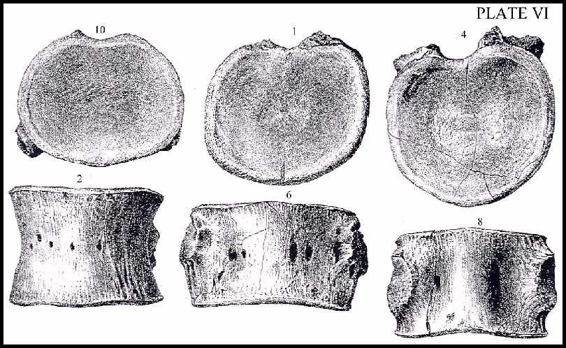
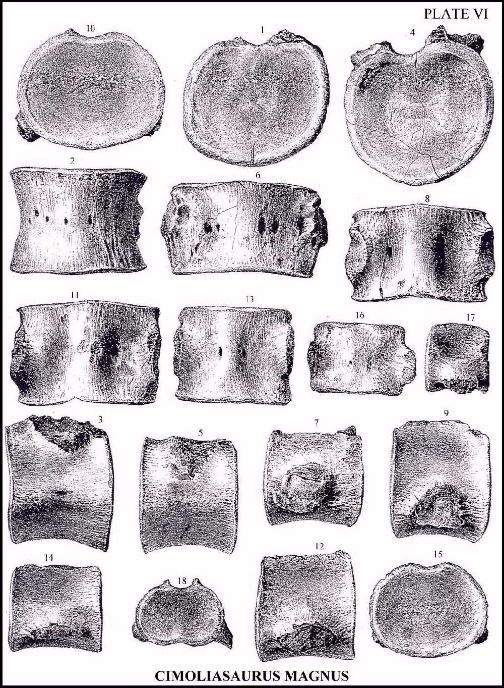
PLATE VI
Vertebræ of Cimoliasaurus magnus, one-half the
diameter of the original specimens. Those from Fig.1 to 15, inclusive were obtained
together, from Freehold, Monmouth County, New Jersey, and apparently belonged to the same
individual. From the collection of 0. R. Willis.
Figs. 1-3. Anterior dorsal vertebra.
Fig. 1. End view, exhibiting a want of symmetry.
Fig. 2. Inferior view.
Fig. 3. Lateral view.
Fig. 4. End view of a large and more anterior dorsal vertebra. It exhibits a
want of symmetry.
Fig. 5. Lateral view of a more posterior dorsal vertebra.
Figs. 6, 7. A vertebra from near the end of the dorsal series.
Fig. 6. Inferior view.
Fig. 7. Lateral view.
Figs. 8, 9. A vertebra, probably the last of the dorsal series.
Fig. 8. Inferior view.
Fig. 9. Lateral view.
Figs. 10-12. A lumbar vertebra.
Fig. 10. End view.
Fig. 11. Inferior view.
Fig. 12. Lateral view.
Figs. 13-15. A posterior lumbar vertebra.
Fig. 13. Inferior view.
Fig. 14. Lateral view.
Fig. 15. End view.
Figs. 16-18. A posterior lumbar vertebra, belonging to the same individual as
Figs. 13-19, Plate V.
Fig. 16. Inferior view.
Fig. 17. Lateral view.
Fig. 18. End view.
|
